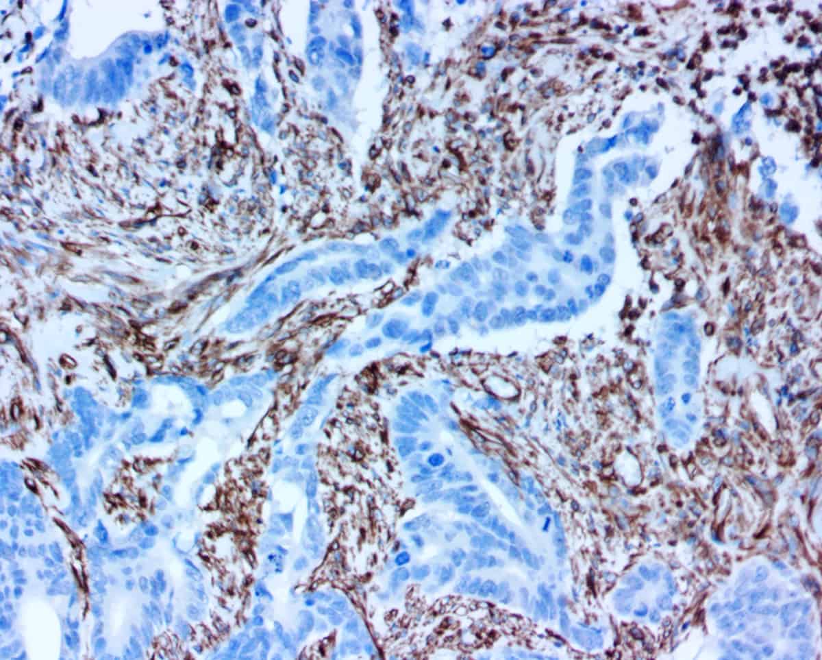
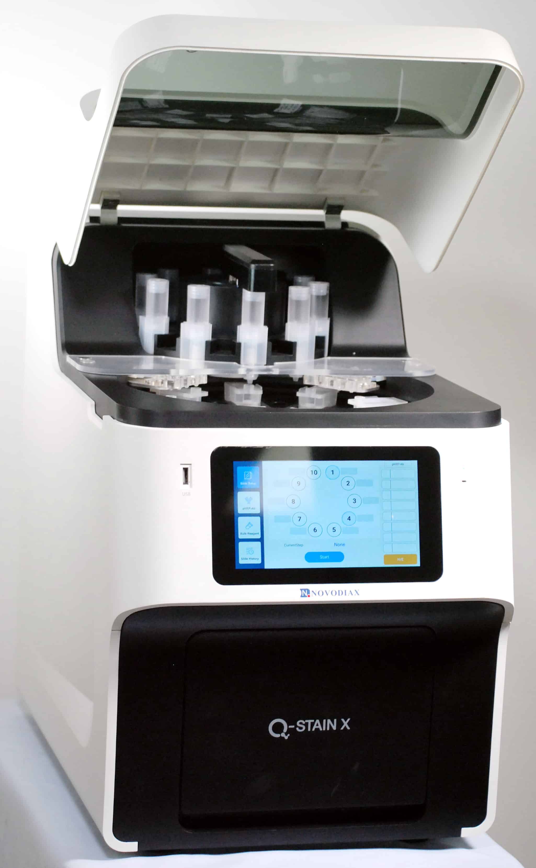


Q-STAIN (QSXTM)-Enh AE1/AE3 antibody (anti human pan-cytokeratin, Clone AE1 and AE3) is intended for laboratory use to qualitatively identify by light microscopy the presence of cytokeratins which are expressed in tissues of epithelial origin, including squamous and glandular epithelial cells, in sections of formalin-fixed, paraffin-embedded tissue sections or frozen tissues using immunohistochemistry (IHC) test methods. The clinical interpretation of any staining or its absence should be complemented by morphological studies using proper controls and should be evaluated within the context of the patient’s clinical history and other diagnostic tests by a qualified pathologist/physician. These reagents have been formulated to ready-to-use concentration and optimized for IHC on specific tissue application use without further dilution.
Summary and Explanation:
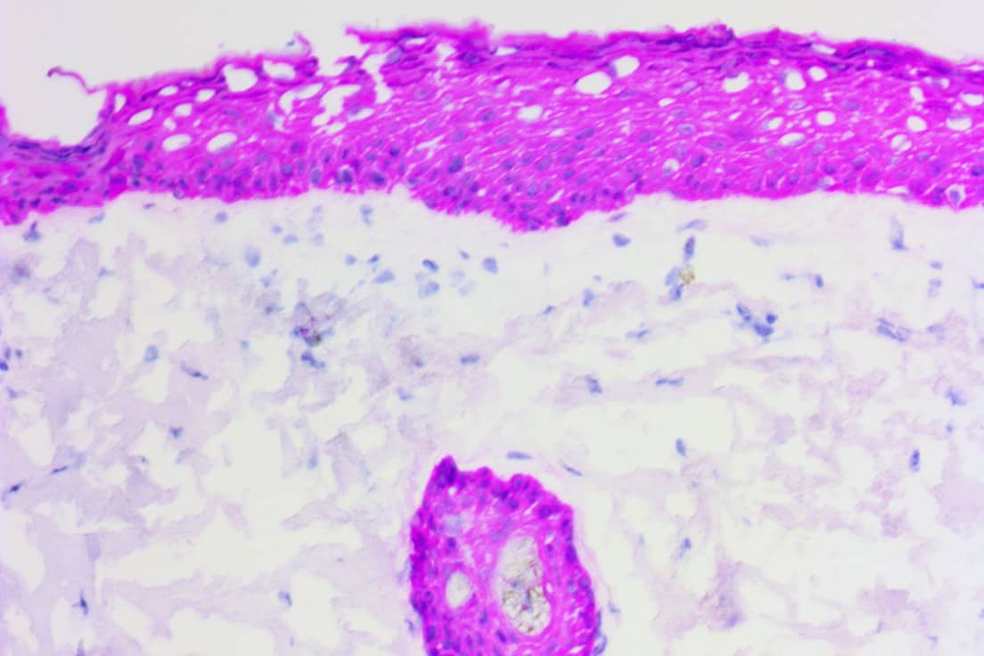
QSX-Enh AE1/AE3 is a ready-to-use antibody reagent (mouse monoclonal antibodies clone AE1 and AE3). Clones AE1 and AE3, always used together as an anti-pan-cytokeratin antibody cocktail, recognize cytokeratins 1-8, 10, 14 -16 and 19 in Western-Blot assay. This test may be applied to individuals suspected of having related forms of cancer. AE1/AE3 antibodies react with cytokeratins which are expressed in tissues of epithelial origin, including squamous and glandular epithelial cells. AE1/AE3 is known to have limited reactivity in hepatocellular carcinomas (HCCs), renal cell carcinomas (RCCs), and pulmonary small cell carcinomas. This cocktail has limited reactivity in some lung cancer specimens and no-to-very poor reactivity with the cytokeratin proteins in epidermal layer of skin.1,2 See Q-STAIN X User’s Guide to learn more about test protocols.


